
Scientists Can Now Find the Brain's Language Network in Any Scan
💡 On August 13, 2026, researchers at Stanford and MIT published a study in Nature Communications confirming that every human brain runs a dedicated language network in the brain - and this network can be identified from any fMRI scan, even when the person is not speaking, reading, or thinking about language at all.
- The study analyzed 1,957 fMRI scans from 1,199 people, the largest dataset of its kind, published in Nature Communications on August 13, 2026.
- Every brain has a language-selective network concentrated in the left frontal and temporal regions, but its precise layout varies from person to person.
- A new method called individualized functional connectomics can locate this network from a resting-state scan, without any language task - a first for the field.
- This could help the estimated 2 million Americans with aphasia: surgeons and therapists may now map language regions even in patients who cannot follow scanner instructions.
- Honest caveat: the automated clustering algorithm can occasionally produce imperfect partitions, and only one connectivity-mapping approach was tested.
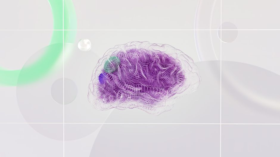
What Scientists Discovered About the Brain's Language Network
When you read this sentence, your brain is not simply activating a general thinking region. A specific, tightly connected set of regions in the left frontal and temporal lobes switches on for language - and this holds for every person who uses a human language. Neuroscientists have hypothesized this for decades, but confirming it precisely required a dataset large enough to be convincing.
Lead author Cory Shain of Stanford University and cognitive neuroscientist Evelina Fedorenko of MIT analyzed 1,957 fMRI scanning sessions from 1,199 individuals, using data collected in Fedorenko's lab between 2007 and 2024. The result, published August 13 in Nature Communications, confirmed that a language network in the brain exists consistently across all participants - and crucially, it is detectable even when no language task is being performed.
Why Was This So Hard to Find Before?
Earlier brain mapping used anatomical coordinates: fixed locations in a standard 3D brain template applied to everyone. The problem is that brains are not identical. Language regions vary in position, size, and shape between individuals. A one-size-fits-all coordinate system often missed or blurred these regions.
A second obstacle was activation. Most language-network mapping required participants to actively read, speak, or listen while inside the scanner. That made it impossible to map language regions in people who cannot follow instructions - including many stroke survivors, children, and patients under sedation.
How Did Researchers Actually Map It?
The new method is called individualized functional connectomics. Instead of looking at where the brain activates in response to a task, researchers tracked which regions' activity rises and falls together over time, even when the person is at rest or doing something unrelated to language, like solving a visual puzzle or listening to music.
The language network emerged from these correlation patterns without prior anatomical assumptions. Each participant's network had its own precise layout, but all followed the same broad plan: left-hemisphere bias, concentrated in frontal and temporal cortex, with an average laterality index of 0.61 (on a scale where 1 is fully left-sided). Shain summarized: "The human brain appears to have a network that's selective and necessary for language, and we can find that network from the activity in the brain alone."
What Does This Mean for People with Language Disorders?
This is where the research moves from interesting to genuinely useful. Approximately 2 million Americans live with aphasia, a language disorder most often caused by stroke. Many cannot follow the verbal instructions used in standard language-activation scans, making pre-surgical brain mapping impossible for them. Individualized connectomics requires no instructions and no task. A resting-state fMRI - the kind a patient can have while lying still - is enough. This opens the door to:
- Pre-surgical language region mapping for patients who cannot follow scanner instructions
- Better-targeted speech therapy based on each patient's individual network layout
- More accurate sensor placement for neural speech prostheses (devices that convert brain signals to speech)
- Identifying optimal targets for brain stimulation treatments
The finding applies beyond stroke: anyone whose language regions need to be located before brain surgery, regardless of ability to cooperate during a scan, could benefit from this approach.
What Does This Tell Us About Language Itself?
For a professional translator or anyone who works with language daily, there is something worth sitting with here: the brain reserves a dedicated circuit for language, distinct from general thinking, memory, or attention. Language is not just another cognitive function layered on top of general intelligence. It has its own address in the brain, and that address is remarkably consistent across cultures, across languages, and across lifetimes.
A related study earlier in the year explored how the bilingual brain organizes two languages within a single grammar engine. These new connectomics findings add another layer: not only does language have a dedicated network, but this network can be precisely located in each individual person, regardless of how many languages they speak.
What Are the Real Limits of This Study - and What's Next?
Shain and Fedorenko are clear about where the method falls short. The automated clustering algorithm that groups correlated brain regions can occasionally produce imperfect results, misassigning a region to the wrong network. The team also tested only one connectivity-mapping approach; other methods might draw the network boundaries differently.
The dataset, while the largest for this question, comes from a single lab. Independent replication across different scanners, populations, and data-collection protocols is needed before clinical use becomes standard. "We can map language networks from any scan" should be understood as "this works consistently in research conditions," not as "this is ready for every hospital next month."
The finding also says nothing about how language is processed - only where. For anyone curious about the intersection of AI and language, a recent post on AI sign language translation shows how machines are learning to handle language in increasingly flexible ways, though connecting that to brain architecture remains speculative for now.
FAQ
What is the brain's language network and where is it?
It is a set of regions in the left frontal and temporal lobes that activates selectively for language: reading, speaking, listening, and even thinking in words. The study confirmed this network exists in all individuals and can be detected from brain activity correlation patterns, even without any language task being performed.
Can this help aphasia patients?
Potentially yes. Aphasia affects roughly 2 million Americans, most often after stroke. Many cannot follow scanner instructions, making standard pre-surgical mapping impossible. Individualized connectomics works from resting-state scans, opening the door to language mapping for patients previously excluded. Clinical application still requires independent validation before becoming routine.
Does everyone have the same language network?
The overall structure is consistent: left-hemisphere bias, concentrated in frontal and temporal regions, with a laterality index averaging 0.61. But the precise layout - which areas are included and how they connect - is unique to each person, much like how human faces follow the same basic plan but no two are identical.
How is this study different from previous brain language research?
Earlier work used anatomical coordinates (fixed positions in a standard template) or required active language tasks (reading, listening). This study uses functional connectivity, tracking how brain regions co-activate even at rest, to find each person's network from within their own brain. It also uses the largest dataset assembled for this question: 1,957 scans from 1,199 participants.
What does this mean for language learning or translation?
Directly, not much yet: the study maps where language lives in the brain, not how learning or translation works. But it reinforces that language is a distinct cognitive system with its own dedicated neural real estate, aligning with the practical reality that language skills - including translation - are something the brain maintains and improves as a specific domain.
Source: News-Medical - Brain scans reveal language-specific network of neurons even during nonlinguistic tasks (2026); The Brighter Side of News - Stanford researchers discover a language-specific network hidden in the human brain (2026)
About the author
Dao Huy (Lucas) is a professional translator working across English, Vietnamese, Chinese, and French, with more than seven years of experience. He follows the frontier of language science not as a neuroscientist, but as someone who works with language every day and finds it genuinely valuable to understand how the brain organizes the work he does for a living. A study showing that language has its own dedicated address in the brain, one consistent regardless of task, culture, or language spoken, is both professionally interesting and personally meaningful.
If you need accurate, certified translation between English and Vietnamese - including technical, software, or IP content - Lucas offers professional services and is happy to provide a quote at daohuy.com.
Written by Dao Huy (Lucas), Vietnamese translator & localization specialist (EN · ZH · FR → Vietnamese). See translation services →